Figure 1.

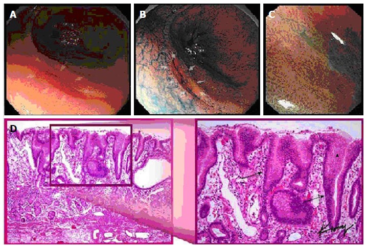
Multiple views of a signet ring cell gastric carcinoma in a single patient: (A) standard white light endoscopy, (B) chromoendoscopy, (C) magnification endoscopy, and (D) histopathology demonstrating elongated gastric glands (arrows) infiltrated with tumor cell (arrowhead).
