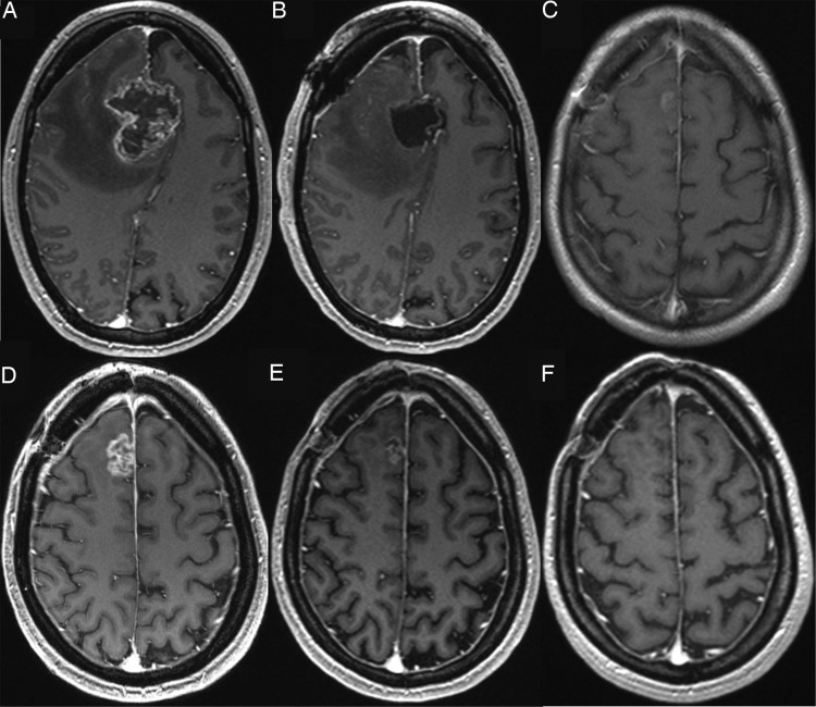
Fig. 1.
A 39-year-old female patient with glioblastoma in the right frontal lobe. Contrast enhanced T1-weighted MRI (A) before and (B) after surgery. (C) First f/u 1 month after completion of radiotherapy shows only minimal enhancement. (D) Enhancement increase of at least 25% appears in the second f/u 4 months after completion of radiochemotherapy. After (E) 7 months the enhancement was rarely visible and disappeared totally after (F) 10 months.

