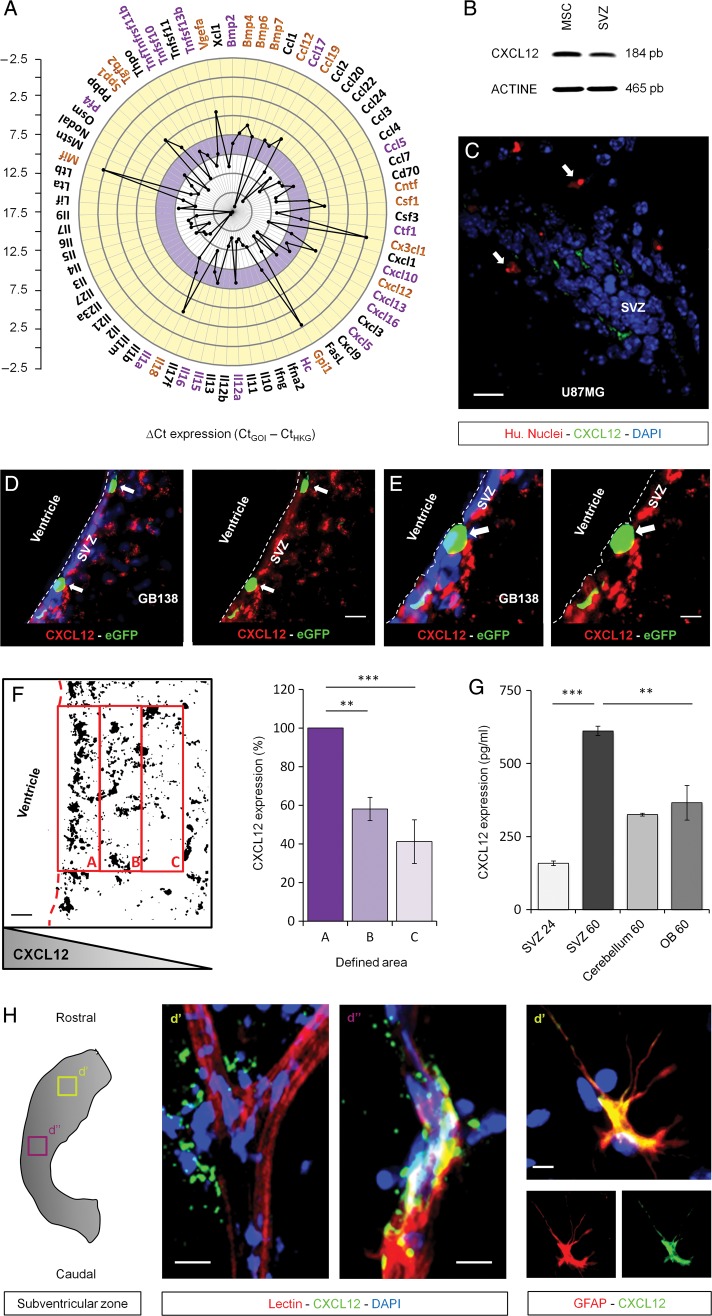
Fig. 2.
Expression of CXCL12 by the subventricular zone (SVZ) environment. (A) RT-qPCR screening notably displayed a high expression level of CXCL12 in the SVZ environment (yellow rim of the graph). (B) This observation was validated by RT-PCR using mesenchymal stem cells (MSC) as a positive control. (C–E) The expression of CXCL12 was next demonstrated on brain coronal sections. This expression was consistent with the presence of U87MG cells (Hu. Nuclei) or GB138 primary cells (eGFP) in the SVZ (white arrows). (F) CXCL12 acquisitions were processed as binary images. The mean intensity, with foreground 255 and background 0, in predefined areas of the SVZ environment (A, B, and C) was calculated. A constant decrease of the CXCL12 expression was observed starting from area A to end at area C, suggesting that CXCL12 is secreted along a decreasing concentration gradient. (G) CXCL12 levels were evaluated by ELISA in conditioned media from SVZ, cerebellum, and olfactory bulb (OB) whole mounts for 24 or 60 hours. (H) CXCL12 was expressed by astrocytes and endothelial cells within the adult SVZ. Immunostaining on organotypic whole mounts showed a closely related expression of CXCL12 (green) with the vasculature and astrocytes (red). SVZ blood vessels and astrocytes were respectively stained using a FITC-coupled lectin or a specific anti-GFAP antibody (red). Cell nuclei were counterstained with DAPI (blue). Scale bars = 20 µm for C and D and 10 µm for E and H. Caption indicates where pictures and materials were taken.

