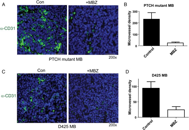
Fig. 4.
MBZ reduced the formation of tumor vessels in the PTCH mutant allograft and D425 xenograft medulloblastoma. (A) The sections of PTCH mutant allograft (PTCH+/−, p53−/−) medulloblastoma (MB) were stained with anti-CD31 antibody and green fluorescent secondary antibody. Nuclei were stained with DAPI (blue). Microvessel density was counted on 200× fields (“hotspot”) in 5 independent slides in each group, graphed in (B). The same frames of green fluorescence pictures were observed with the Texas Red fluorescence filter, in order to rule out possible autofluorescence. All pictures were taken with the setting of 800 msec exposure to green fluorescence and 200 msec to DAPI channel. All scale bars are 30 μm. (C, D) Similar staining and measurement were performed with D425 medulloblastoma xenografts.

