Fig. 3.
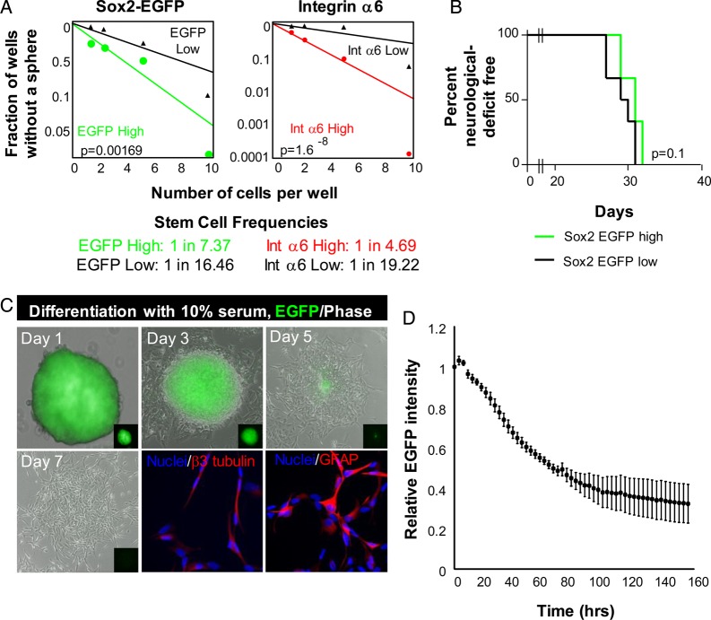
Sox2–EGFP-positive glioma cells display some functional characteristics of cancer stem cells. (A) In vitro limiting-dilution assay to determine self-renewal in Sox2–EGFP-high cells sorted for EGFP and integrin α6 expression. (B) Kaplan-Meier survival curve demonstrates no difference in tumor initiation between Sox2–EGFP-high (green line) and -low (black line) glioma cells. (C) Representative images of tumorsphere-forming Sox2–EGFP-high glioma cells in the absence or presence of the prodifferentiation agent fetal bovine serum (FBS). Sox2–EGFP-high glioma cells lost EGFP expression in the presence of 10% FBS and gave rise to neural (β3 tubulin-positive) and astrocyte (GFAP-positive) lineages. (D) Quantification of EGFP signal over one week of differentiation in the presence of 10% FBS. Graph is representative of quadruplicate samples.

