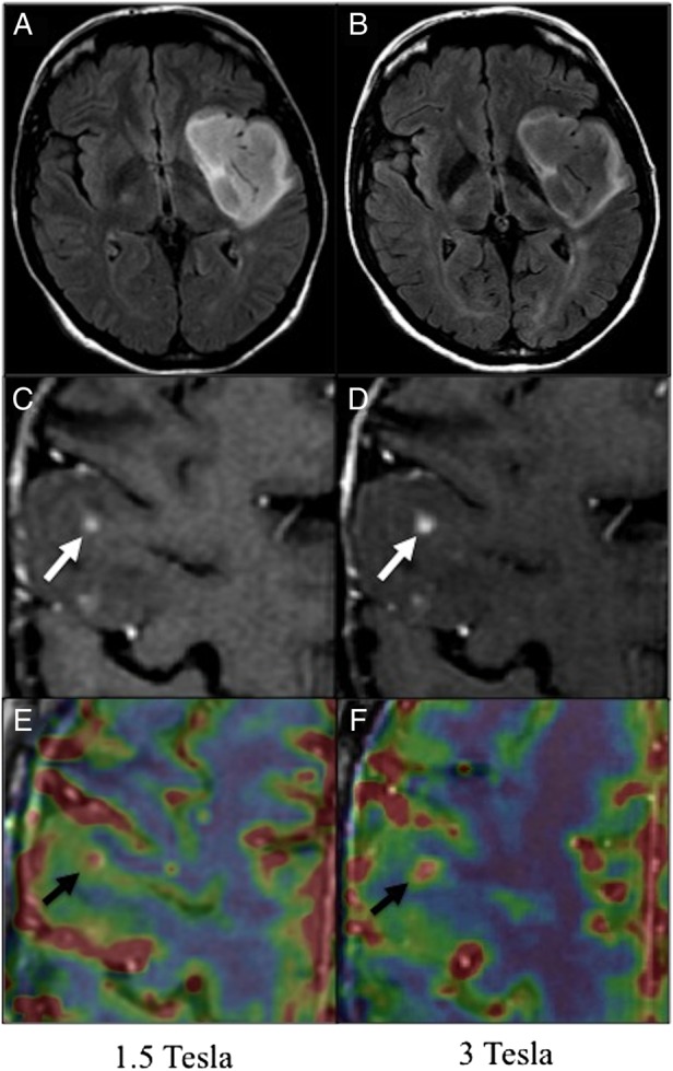
Fig. 1.
Patients with glioma at 1.5T (left column) and 3T (right column). A and B. Axial FLAIR sequence at 1.5T (A) and 3T (B) of a 51-year-old woman with a left frontal glioma (WHO grade II) treated with radiation therapy 9 years before. Time interval between examinations: 3 days. C, D, E and F. Post-contrast 3D T1-w (C, D), and perfusion images (E, F) of a 56-year-old patient with a progressing grade II oligoastrocytoma treated 10 years before with surgery and radiation therapy, showing nodular contrast enhancement (white arrows) at 1.5 (C) and 3-T (D). Focal rCBV increase visible as a “hot spot” (high values in red, low values in blue) on co-registered perfusion parametric map and post-contrast 3D T1-w images (black arrows) at 1.5 (E) and 3T (F). Time interval between examinations: 7 days.