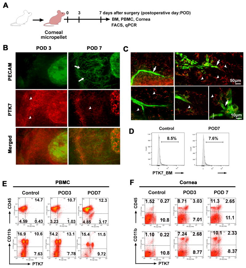
Figure 1. PTK7+ cells recruit to the cornea after VEGF-A-induced neovascularization.
A, Schematic drawing illustrates the VEGF-A micropellet-induced corneal angiogenesis experiment. To induce corneal neovascularization VEGF-A (160 μg) micropellets were inserted into Balb/c mouse corneas (n=5). Corneas were harvested before micropellet implantation (day 0) and at postoperative day (POD) 3 and 7. B and C, Corneas were isolated at postoperative day (POD) 3 and 7, immunohistochemically stained for PECAM-1 (green) and PTK7 (red), and then examined by epifluorescence microscopy (× 200) (B) and confocal microscopy (C); white arrows indicate new vessels, arrowheads indicate PTK7+ cells. D, Representative flow cytometry histogram showing the percentage of PTK7+ cells in the bone marrow (BM) before (Control) and 7 days after micropellet implantation (POD7). E and F, Representative flow cytometry dot plots showing PTK7+CD45+ (upper row) and PTK7+CD11b+ (lower row) cells in peripheral blood mononuclear cells (PBMCs) (E) and cornea (F) before micropellet implantation (Control), and 3 (POD3) and 7 days (POD7) after implantation.

