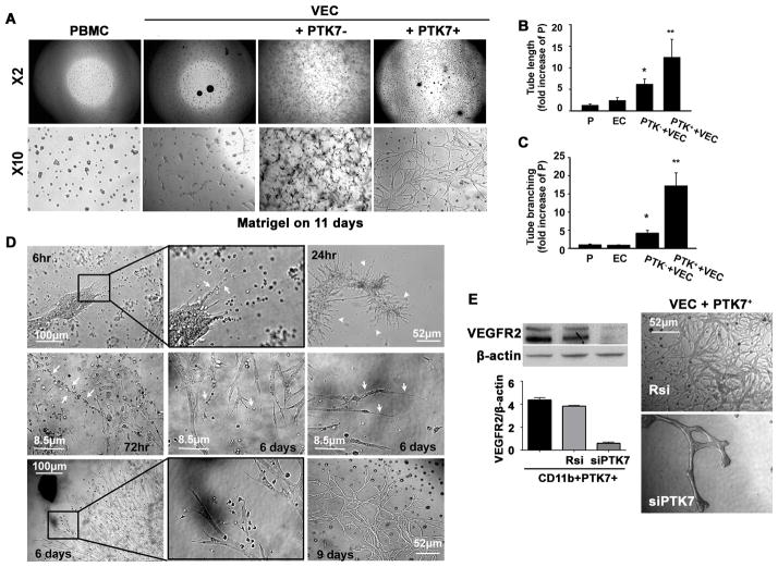
Figure 4. PTK7+ mononuclear cells facilitate vessel growth and stability in vitro.
A, The tube formation assay was performed using a mouse vascular endothelial cell (VEC) line (MS1) and PTK7+ or PTK7− CD11b+ peripheral blood mononuclear cells (PBMCs;). Images were obtained using an inverted microscope at 11 days later (×2 and ×10: objective lens magnification). B and C, Five high-magnification (200×) pictures for each condition were taken, and the tube length (B), and branching (C) were measured using image analysis software. *p < 0.05 vs. PBMC, **p < 0.01 vs. PBMC. (VEC: vascular endothelial cell, PTK7−: PTK7−CD11b+, PTK7+: PTK7+CD11b+). The results are mean ± SD from three independent experiments. D, VECs were co-cultured with PTK7+ or PTK7− PBMCs and images were taken as early as 6 h and up to 9 days after starting the coculture. White arrows in the upper center panel (6 hours) indicate palisading PBMCs (black box, upper row). The white arrowheads in the upper right panel indicate new vessel buds growing from clusters of endothelial cells at 24 hours. Linear or palisading PBMCs were found near the growing vascular network (white arrows in the middle left panel; 72 hours). Cells with hair-like long processes were observed at the end of the blood vessels (white arrows in the middle center and middle right panel). Macrophage-like cells were linked to growing endothelial buds and the tips of the growing tubes (black box, lower row). A stable vascular network was observed up to 9 days after culture. E, PTK7+CD11b+ PBMCs were transfected with random siRNA (Rsi) or siVEGFR2 (siR2) and VEGFR2 expression levels was determined by immunoblot at 72 hours after transfection. Then, VEGFR2 knock down PTK7+CD11b+ cells were cultured with VECs for the tube formation assay. 14 days later we observed less tube formation in VECs when co-cultured with VEGFR2 knock down cells.

