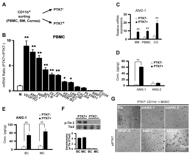
Figure 5. PTK7+ cells enhance vascular stability through angiopoietin-1.
A, A schematic drawing illustrates experiments for Figure 5A–E. B, Real-time qPCR analysis showing the expression of VEC markers in PTK7+CD11b+ and PTK7−CD11b+ cells. Comparison of the expression of N: GAPDH to the expression of empty pellets of vascular endothelial cell growth factor receptor (VEGFR) 2, Angiopoietin 1 (ANG-1), Neurophilin 1 (NRP1), Apelin (APLN), Delta-like 4 (DLL4), Ephrine B2 (EPHRIN B2), VEGFR1, unc-5-homolog B (UNC5B), CXCR4, plexin D1 (PLXND1), Neuropilin-2 (NRP-2), and CD34 was done. C, Three days after pellet insertion PTK7+CD11b+ and PTK7−CD11b+ cells were sorted by FACS from the bone marrow (BM), peripheral blood mononuclear cells (PBMC), and cornea. mRNA expression of ANG-1 was assessed using real-time qPCR. The fold increase compared to non-operated control mice was measured. D, Protein expression of ANG-1 and ANG-2 of in vitro cultured PTK7−CD11b+ and PTK7+CD11b+cells measured by ELISA after 24 hours of VEGF-A (40ng/ml) treatment. E and F, 5 × 10⁴ PTK7+CD11b+ cells were cultured with 2 × 10⁵ vascular endothelial cells (VECs) using a Boyden chamber (BC) or a mixed co-culture (MC). ANG-1 protein concentration (E) and phospho-Tie2 and total Tie2 (F) were measured by ELISA or immunoblot, respectively, 24 hours later. G, PTK7+CD11b+ cells were sorted and transfected with random siRNA (Rsi), siANG-1, siANG-2, or siPTK7. Then, cells were co-cultured with VECs in a matrigel for 5 days. As indicated 2 co-culture conditions of siPTK7 transfected cells and VECs were treated with 40ng/ml of ANG-1 or ANG-2 every 24 hrs. All experiments were repeated four times. Data are expressed as the mean ± standard deviation. *p < 0.05; **p < 0.01.

