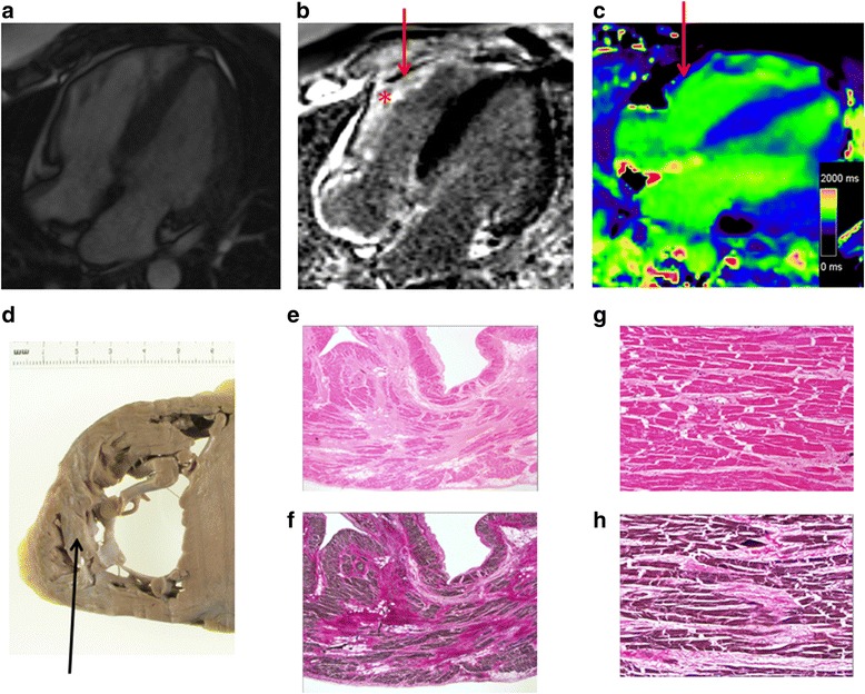
Fig. 4.

Myocardial fibrosis on CMR and post-mortem specimens from a patient with ALMS. a-c) 4 chamber CMR imaging; a cine, b late gadolinium enhancement and c T1 mapping. Note the extensive LGE in the RV (*) with corresponding low T1 on the post-contrast MOLLI. d Corresponding macroscopic image from autopsy of the RV with strands of sub-endocardial fibrosis seen as pallor within the RV (black arrow). e-f Corresponding low power views of part of the free wall of the right ventricle demonstrating patchy swathes of interstitial fibrosis replacing myocardiocytes with more subtle pericellular fibrous expansion. The fibrous tissue stains pink with H&E and red with EHVG (H&E & EHVG, original magnification X1.25). g-h Comparable intermediate magnification photomicrographs of the free wall of the right ventricle depicting more dispersed interstitial fibrosis enclaving groups of myocytes and focally insinuating between individual muscle cells. The fibrous tissue stains pink with H&E and red with EHVG (H&E stain, original magnification X1.25)
