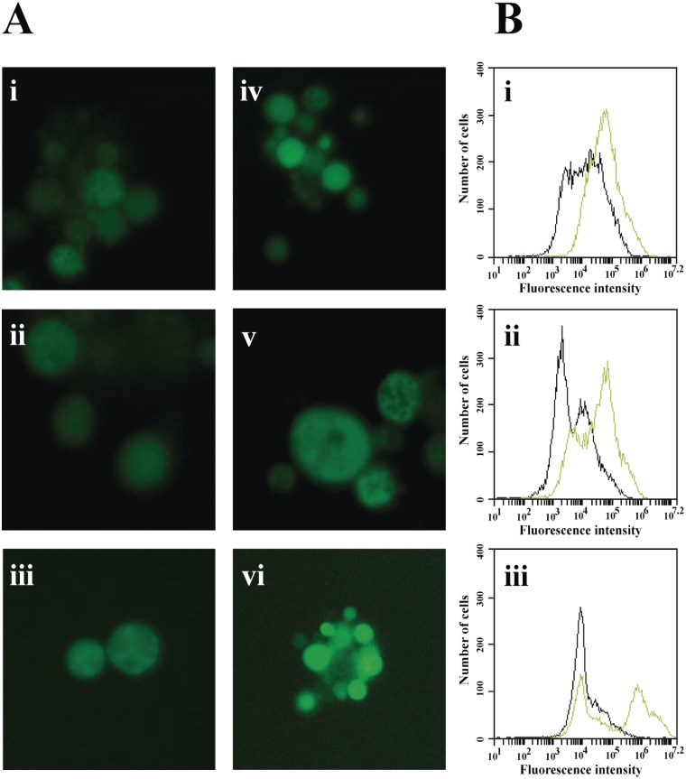
Fig 5. Formation of ROS by TSC-C.
(A) Fluorescence microscopy of P. lutzii yeast cells stained with 2`,7`-dichlorofluorescein diacetate. Yeast cells were grown in the absence of TSC-C for i) 4 h, ii) 8 h and iii) 12 h and in the presence of TSC-C for iv) 4 h, v) 8 h and vi) 12 h. (B) Flow cytometry analysis of yeast cells grown in the absence or in the presence of TSC-C. The cells were monitored for i) 4 h, ii) 8 h and iii) 12 h stained with 2`,7`-dichlorofluorescein diacetate. Black histograms represent control yeast cells, and green histograms represent yeast cells treated with TSC-C.

