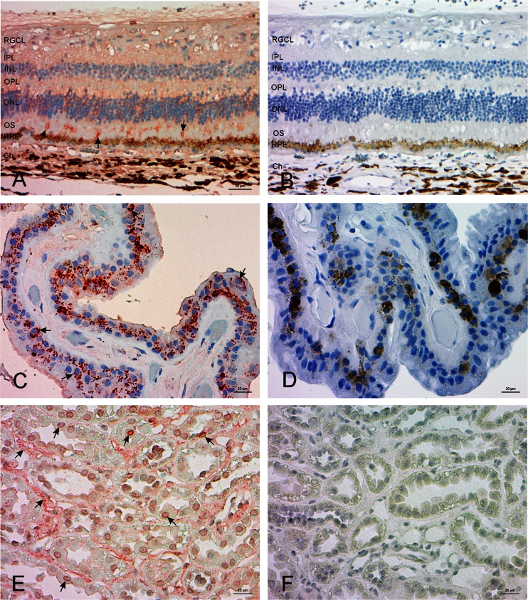
Fig. 7.
Expression of cytoplasmic Ang(1–7) in the human retina and ciliary body. A weak immunostaining of Ang(1–7) was found in the inner and outer nuclear and plexiform layers of retina a In the ciliary body there was a cytoplasmic stain in the non-pigmented and pigmented epithelial cells (c). Magnification 200×. A human kidney sample is shown in e. Magnification 400×. In all figures, red–brown color indicates staining of Ang (1–7), see black arrows. b, d, f Show negative staining samples without labelling antibody. RGCL = retinal ganglion cell layer, IPL = inner plexiform layer, INL = inner nuclear layer, OPL = outer plexiform layer, ONL = outer nuclear layer, OS = outer segment, RPE = retinal pigment epithelium, Ch = choroidea

