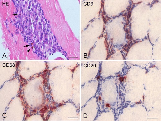
Fig. 1.

Muscle biopsy of patient #1. a Hematoxylin and eosin (HE) stain showing an endomysial inflammatory cell infiltrate with eosinophils (arrows). b (CD3) T lymphocytes with invasion of muscle fibers. c (CD68) macrophages. d (CD20) B lymphocytes. Scale bars, 40 μM
