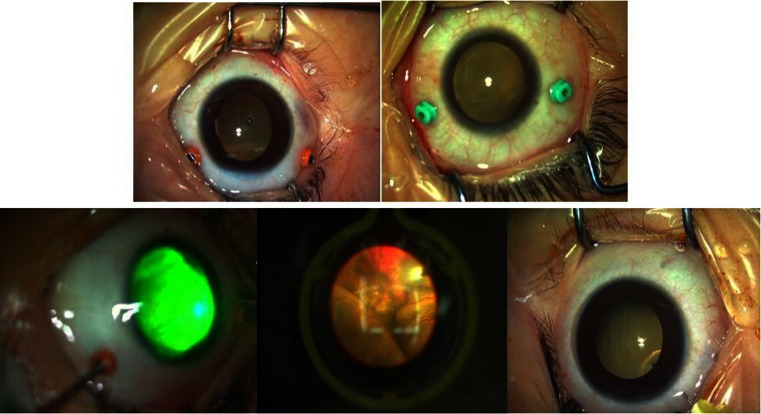
Fig. 1.
Illustration of the surgical procedure of a 4-year-old boy with stage 3A Coats’ disease (OS). Top left Two 23-gauge incisions made 3 mm posterior to the corneal limbus. Top right Two 25-gauge incisions made 3 mm posterior to the corneal limbus. Bottom left Endolaser ablation of telangiectasias. Bottom center Endolaser in the noncontact wide-field viewing system. Bottom right Anti-vascular endothelial growth factor (VEGF) injection

