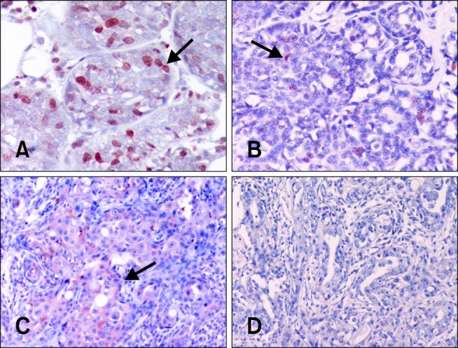
Fig. 2. Immunohistochemical staining for Ki-67 and B-cell lymphoma 2 (Bcl-2). (A) Pre-treatment Ki-67-positive cells (arrow). (B) Post-treatment Ki-67-positive cells (arrow). (C) Pre-treatment Bcl-2-positive cells (arrow). (D) Post-treatment Bcl-2-positive cells. Immunoperoxidase technique with Mayer's haematoxylin counterstaining. Magnification: 240× (A), 180× (B and C), 140× (D).

