Figure 1.

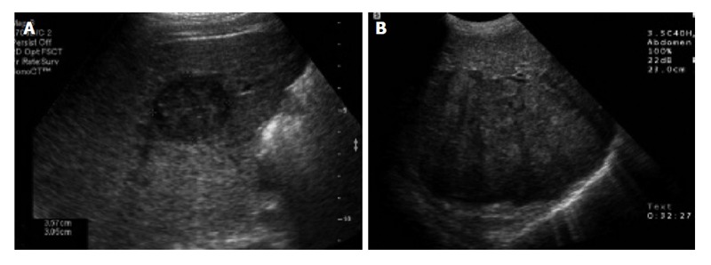
Abdominal ultrasound of the liver. A: Transverse sonogram shows a small, 3 cm, hypoechoic mass in the right lobe of the liver; B: Transverse sonogram shows a heterogeneous large mass in the right lobe of the liver[47].

Abdominal ultrasound of the liver. A: Transverse sonogram shows a small, 3 cm, hypoechoic mass in the right lobe of the liver; B: Transverse sonogram shows a heterogeneous large mass in the right lobe of the liver[47].