
Figure 3.
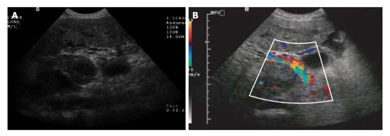
Colour Doppler ultrasound of the liver. A: Transverse sonogram shows portal vein thrombus; B: Transverse colour Doppler sonogram of the right upper quadrant shows heterogeneous flow within the tumour thrombus[47].
Official websites use .gov
A
.gov website belongs to an official
government organization in the United States.
Secure .gov websites use HTTPS
A lock (
) or https:// means you've safely
connected to the .gov website. Share sensitive
information only on official, secure websites.
Colour Doppler ultrasound of the liver. A: Transverse sonogram shows portal vein thrombus; B: Transverse colour Doppler sonogram of the right upper quadrant shows heterogeneous flow within the tumour thrombus[47].