Figure 3.
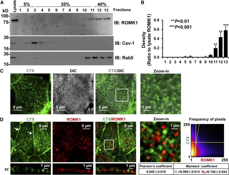
ROMK1 channels are not located in lipid rafts. (A) Sucrose gradient experiments showed that ROMK1 is located in nonlipid raft regions. Caveolin-1 (Cav-1) was used as a control protein that is known to be located in lipid rafts, whereas Rab5 was used as a control protein that is known to be located in nonlipid raft membranes. IB, immunoblotting. (B) Summary plots of four sucrose gradient experiments. (C) Confocal microscopy fluorescent image merged with a DIC image shows that CTX (green) is mainly located in microvilli (microvilli were visualized through DIC imaging). (D) Confocal microscopy shows that ROMK1 (red) is not located in lipid rafts probed by fluorescence-tagged CTX (green). Here and in other figures, all confocal microscopy xy optical sections were taken near the apical membrane of mpkCCDc14 cells, which is evidenced by tight junctions (white arrows); white rectangular boxes indicate zoomed-in areas shown in the Zoom-in panels. Frequency of pixels was analyzed with the ImageJ program, and the images represent data from at least four separate experiments that showed consistent results. All confocal microscopy images in this study were taken using the same parameter settings, including gain, contrast, and pinhole. DIC, differential interference contrast.

