. 2015 Jun 15;39(3):188–197. doi: 10.4093/dmj.2015.39.3.188

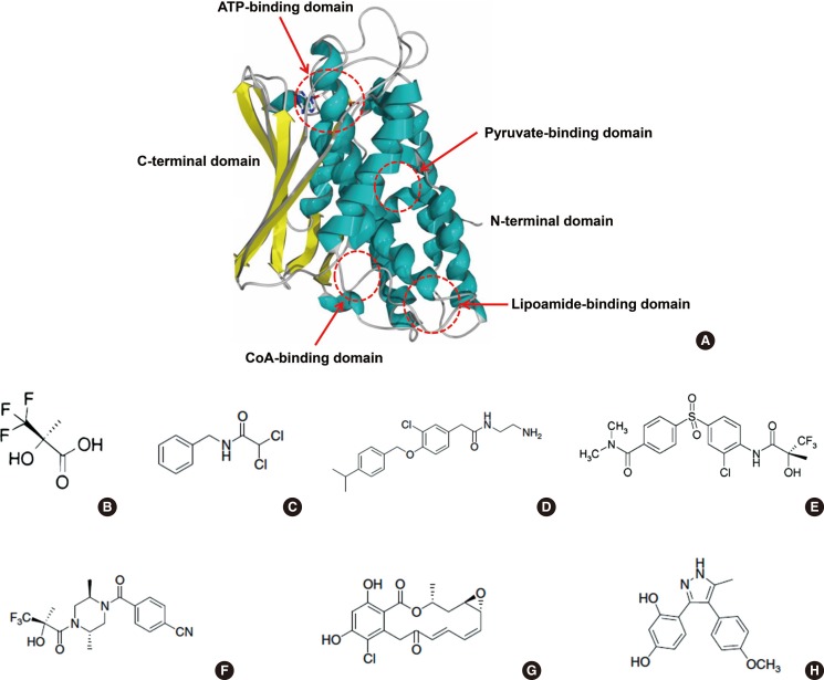
Fig. 4. Allosteric binding domains and substrate binding domains on the pyruvate dehydrogenase kinases (PDKs) (A) and PDK inhibitors (B-H). (A) Protein structure of PDK2 monomer and its allosteric regulator binding sites and substrate binding domains; two allosteric binding domains (pyruvate-binding domain and CoA-binding domain) and one substrate binding domain (lipoamide-binding domain) are located on the N-terminal domain and another substrate binding domain (ATP-binding domain) is located on the C-terminal domain. (B, C) Inhibitors of the pyruvate-binding domain (B, (R)-trifluoro-2-hydroxy-2-methylpropionic acid; C, N-benzyl-2,2-dichloroacetamide). (D) Pfz3, inhibitor of the coenzyme A-binding site. (E, F) Inhibitors of the lipoamide-binding domain (E, AZD7545; F, Nov3r). (G, H) Inhibitors of the nucleotide-binding domain (G, radicicol; H, M77976).

Fig. 4