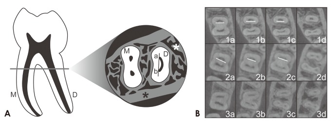
Fig. 2. Curved canal in the cross-sectional image of the distal root in the mandibular first molar. A. Left: a mandibular first molar with (M) mesial and (D) distal roots. Right: cross-sectional image of a mandibular first molar as seen in an axial-plane CBCT view. (M) mesial root, (D) distal root, (*), cortical and trabecular bone structure. When a straight line is drawn in the distal ribbon-shaped canal, it only comes in contact with the (a) buccal and (b) lingual ends of the canal. B. Cone-beam computed tomography cross-sectional images. 1a-c: curved distal canals in cross-sectional images, 1d: an oval canal, 2a and b: curved distal canals in cross-sectional images, 2c: a canal starting to divide, 2d: a fully divided canal, 3a-d: the absence of a curved distal canal in cross-sectional images.