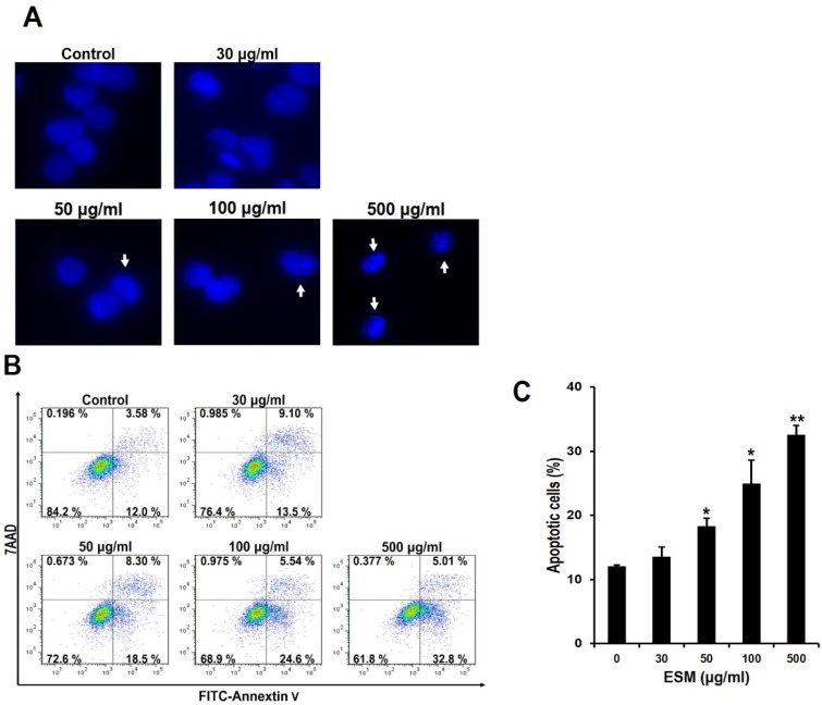
Figure 2.
ESM induces apoptosis in K562 cells. After the K562 cells were treated with the indicated concentrations of ESM for 24 h under serum free conditions, cells were stained with DAPI. The morphological features of the nuclei were then determined by fluorescent microscope (A). Under the same conditions, after the K562 cells were stained with Annexin V-FITC/7AAD, the Annexin V-FITC and/or 7AAD positive cells were detected by flow cytometry (B); Quantitative analysis of the Annexin V-FITC positive cells was then carried out (C); Data are presented as the mean ± SEM (n = 3). ** P < 0.01 and * P < 0.05 compared with control.

