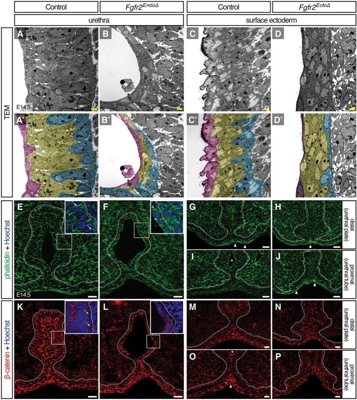
Fig. 5.
Deletion of Fgfr2 disrupts epithelial cell shape, cytoskeletal organization, and cell adhesion. (A-D′) Ultrastructural analysis by TEM reveals aberrant cell shapes in Fgfr2– genital tubercle epithelia; mutant basal cells fail to elongate perpendicular to the basement membrane in both Fgfr2EndoΔ and Fgfr2EctoΔ mutant epithelia. Diminished stratification is evident in the Fgfr2EndoΔ urethra. Cells adjacent to basement membrane are pseudocolored cyan, those neighboring the lumen are magenta and intervening cells are yellow. Scale bars: 2 μm. (E-P) Fluorescent micrographs of control, Fgfr2EndoΔ and Fgfr2EctoΔ genital tubercles stained with phalloidin (E-J) or immunolabeled with an anti-β-catenin antibody (K-P). Note the long cell cortices stained with F-actin in E (white arrows) and short, hatched phalloidin domains in F (arrowheads). Punctate, basolateral foci of β-catenin are evident in control basal urethral cells (yellow arrows) but were not detected in Fgfr2EndoΔ mutants. In controls, the ventral seam shows enriched actin stress fibers (white triangles) distally and increased β-catenin staining (yellow triangles) proximally. Elevated abundance of F-actin fibers is coincident with static levels of β-catenin intensity along the proximodistal ventral midline of Fgfr2EctoΔ mutants. Asterisks mark urethral lumen, dotted lines outline the urethral and surface epithelia. Scale bars: 20 μm.

