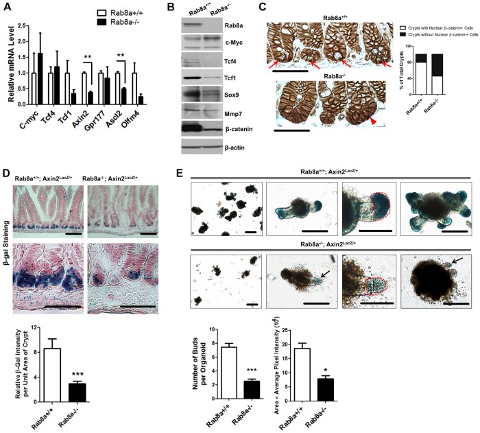
Fig. 3.
Rab8a deletion impairs canonical Wnt signaling in intestines. (A) Quantitative RT-PCR showed reduced Tcf1, Olfm4, Axin2 and Ascl2 expression in Rab8a−/− intestines. (B) Western blots showed reduced Tcf1, Tcf4, Sox9 and β-catenin levels in Rab8a−/− intestines. (C) Immunohistochemistry for β-catenin showed a reduced number of crypts with nuclear β-catenin+ cells (arrows). Note that only crypts with detectable Paneth cells were scored for their positive or negative inclusion of nuclear β-catenin (n=50 for each genotype). (D) β-Gal staining of mouse small intestines showed a significant reduction of Axin2 reporter activity in crypts in Axin2lacZ/+;Rab8a−/− mice. Thirty continuous crypts were analyzed in each section of independent wild-type and knockout mice. (E) β-Gal-stained Axin2lacZ/+ and Axin2lacZ/+;Rab8a−/− intestinal organoids showed significantly reduced bud number and size in the absence of Rab8a. Images were taken at day 10 after crypt plating. Arrow points to a small bud. Twenty organoids of each genotype were quantified for β-gal-stained bud areas (circled in red). *P<0.05, **P<0.01, ***P<0.001. Scale bars: 10 μm in C,D; 15 μm in E.

