Fig. 7.
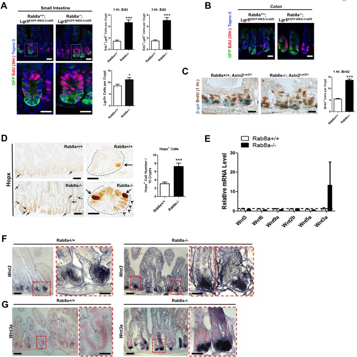
Adaptive changes in Rab8a−/− crypts. (A) EdU labeling (1 and 3 h, red) of mouse small intestine showed a significant increase in proliferative Lgr5+ cells in Rab8a−/−;Lgr5EGFP−IRES−CreERT2 mice. The total number of Lgr5+ cells was also increased in Rab8a−/− crypts. Around 45 crypts that contained Lgr5+ cells were analyzed in each tissue section of independent Lgr5EGFP−IRES−CreERT2 or Rab8a−/−;Lgr5EGFP−IRES−CreERT2 mice. (B) EdU labeling of the colon showed an increase in proliferative Lgr5+ cells in Rab8a−/−;Lgr5EGFP−IRES−CreERT2 mice. (C) Co-staining of BrdU (1 h, brown) and β-gal (blue) showed increased BrdU+ cells in Axin2lacZ/+;Rab8a−/− intestines, despite decreased Axin2 reporter activity in the same crypts. One hundred continuous crypts were analyzed in each section of independent Rab8a+/+ and Rab8a−/− mice. (D) Rab8a−/− intestines contained more cells with the strongest level of Hopx immunoreactivity (arrows). Arrowheads point to cells with moderately higher immunoreactivity than in wild type. One hundred continuous crypts were analyzed in each section of independent Rab8a+/+ and Rab8a−/− mice. (E) Quantitative RT-PCR detected increased Wnt3a levels in Rab8a−/− intestines (n=3 for each genotype). (F,G) RNA in situ hybridization to detect Wnt3 (F) or Wnt3a (G), showing ectopic activation of Wnt3a in Rab8a−/− crypts, whereas Wnt3 was largely unaffected. *P<0.05, ***P<0.001. Scale bars: 10 µm.

