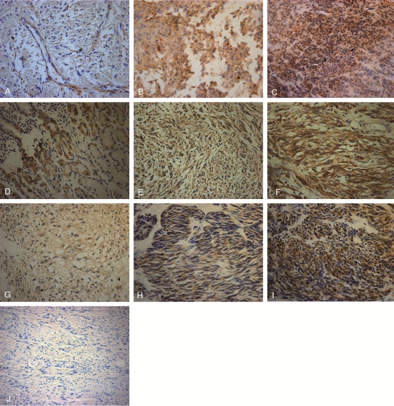
Figure 1.

Representative immunohistochemical staining images of MMP-9, COX-2 and VEGF. Expression of MMP-9, COX-2 and VEGF in GIST tissues was detected by immunohistochemistry. Representative immunohistochemical results were shown. Magnification: × 400. Cells with brown articles were defined as positive. The overall degree of staining was defined as follows: negative staining (-), score 0; weak positive staining (+), score 1-3; positive staining (++), score 4-6; strong positive staining (+++), score 7-9. A. MMP-9 weak positive staining (+); B. MMP-9 positive staining (++); C. MMP-9 strong positive staining (+++); D. COX-2 weak positive staining (+); E. COX-2 positive staining (++); F. COX-2 strong positive staining (+++); G. VEGF weak positive staining (+); H. VEGF positive staining (++); I. VEGF strong positive staining (+++); J. Negative control.
