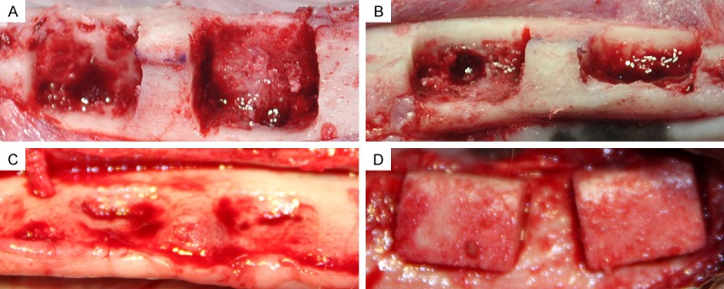
Figure 2.

A and B. (Buccal view and occlusal view, respectively) Standardized box-type defects (9 mm in height from the crestal bone, 6 mm in depth from the surface of the buccal bone, and 12 mm in width mesiodistally; distance between defects is 4 mm) were surgically created at the buccal aspect of the alveolar ridge after removing teeth from the mandible. C. A chronic-type defect was achieved after eight weeks of submerged healing (occlusal view). D. After defect reshaping, nHA/coral blocks were implanted into defect sites (buccal view).
