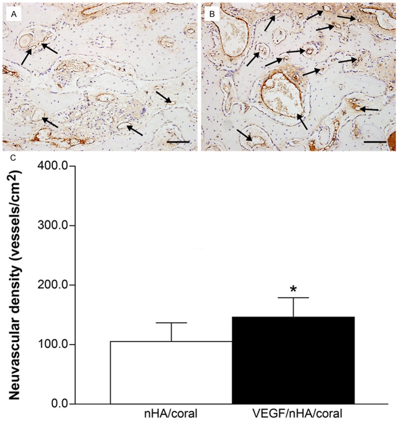
Figure 6.

(A and B) Immunohistological staining for von Willebrand factor of decalcified tissues treated with block grafting. Representative images of nHA/coral section (A) and VEGF/nHA/coral section (B) show circular, dark brown vessels (black arrows) in the scaffolds. VEGF/nHA/coral samples had higher neovascular densities compared to nHA/coral samples. Bar: 100 μm. (C) The blood vessel densities were calculated from the immunohistochemical staining. The local delivery of VEGF promoted the formation of new blood vessels. Columns show mean values, and error bars represent the corresponding standard deviations (n = 8), *P < 0.05.
