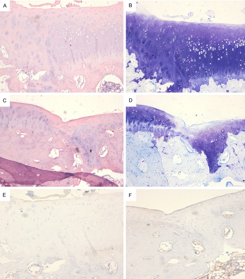
Figure 1.

A: BMSCs group, at 16 weeks after surgery, chondral cells in the repaired tissues lied in the cartilage lacunae, the newly generated cartilages were similar to surrounding normal cartilages and there was no space between surrounding with cartilages/osteochondral grafts and newly generated cartilages. B: At 16 weeks after surgery, the chondral cells located in the cartilage lacunae. Toluidine blue staining performed at 16 weeks after surgery showed a blurry borderline between newly generated cartilage and sub-chondral bone, suggesting a good integration. The newly generated chondral cells showed a close arrangement in the lacuna, but were smaller than normal chondral cells, and the cells in fibrous tissues were larger than normal ones. C and D: In control group, at 16 weeks after surgery, chondral cells in the repaired tissues showed scattering distribution in crumb, and the repaired tissues were only 1/2 of normal or repaired by fibrous-chondral tissues. E: Immunohistochemistry showed the type II collagen was brown. In BMSCs group, at 16 weeks after surgery, the interspace, transplanted with allogenic BMSCs, showed similar staining as in normal cartilage, except for slightly thicker collagens, while the matrix staining was more obvious as compared to normal cartilage. F: At week 16, the chondral cells showed a scattering distribution around in the matrix and an irregular arrangement.
