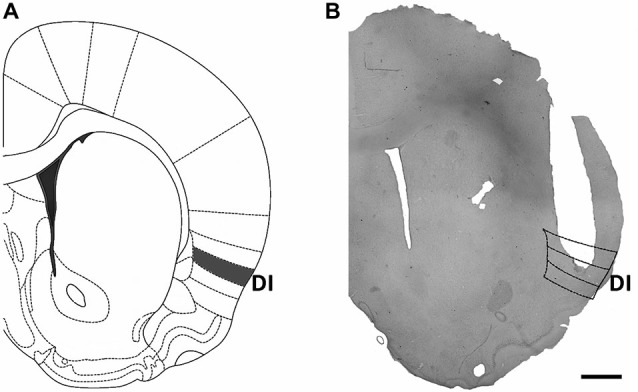
Figure 1.

Location of cannula implants within the insular cortex. (A) Scheme of a brain coronal section showing the dysgranular (gustatory) area of the insular cortex, which was targeted by cannula implants. (B) Microphotograph of a representative cannula implant into the dysgranular (taste) area of the insular cortex. Scale bar: 1 mm.
