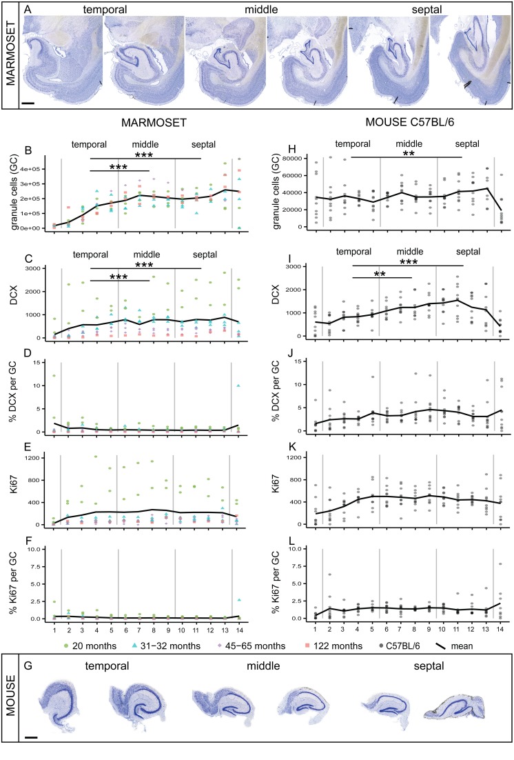
FIGURE 1.
Comparison of septo-temporal gradients in marmoset and mouse.(A) Giemsa-stained coronal sections from the temporal to the septal pool of a marmoset hippocampus (distance between sections 960 μm). Significant gradients along the septo-temporal axis are found in morphed numbers of granule cells (GC, B) and DCX (C). Proliferating cells (Ki67, E) and normalized neurogenesis-related cell numbers expressed as a percentage of local granule cells (% DCX per GC, D; and % Ki67 per GC, F) are evenly distributed along the longitudinal axis. (G) Coronal sections of the matrix-embedded, straightened hippocampus of a C57BL/6 mouse (distance between sections 120 μm). Similar to the marmoset hippocampus, a significant septo-temporal gradient is apparent for granule cells (H) and DCX+ cells (I), but not for the other cell numbers (J–L). All stereologically assessed cell numbers are morphed to 14 virtual sections, section 1 and 14 were excluded from the statistical analysis. Numbers for marmosets are color-coded for individual ages, all C57BL/6 (N = 10) are 14 weeks of age. For exact p-values see result section. Scale bar (A): 1 mm; (G): 500 μm.

