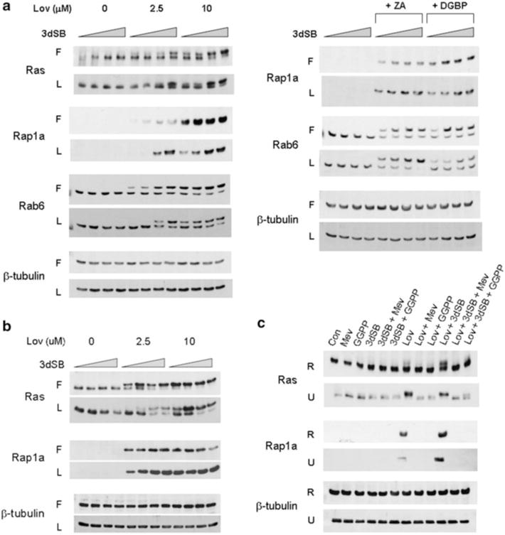
Fig. 3.

3dSB potentiates lovastatin-induced decrease in protein prenylation. a RPMI-8226 or b U266 cells were incubated in the presence or absence of 3dSB ± IBP inhibitor (Lov lovastatin, ZA zoledronic acid (50 μM) or DGBP digeranyl bisphosphonate (2.5 μM)) in either standard FCS (F) or LPDS (L) for 24 h. The wedge indicates increasing concentrations of 3dSB (0, 10, 50, 100 nM for RPMI-8226 cells and 0, 0.1, 0.5, 1 μM for U266 cells). c Lovastatin- or lovastatin + 3dSB-induced decrease in protein prenylation is prevented by co-incubation with select isoprenoid intermediates. RPMI-8226 (R) and U266 (U) cells were incubated for 24 h in media containing standard FCS in the presence or absence of lovastatin (10 μM), 3dSB (50 nM for RPMI-8226 and 1 μM for U266), mevalonate (1 mM), or GGPP (10 μM). Western blot analysis of Ras (more slowly migrating band represents unmodified Ras), Rap1a (antibody detects only unmodified Rap1a), Rab6 (more slowly migrating band represents unmodified Rab6) and β-tubulin (as a loading control) was performed. Data are representative of at least two independent experiments
