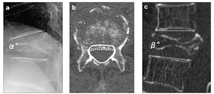
Fig. (3).

Radiographic parameters: (a) “local kyphotic angle” (α°) measured as the angle between the lower and upper endplates of the uninvolved vertebrae adjusted cephalic and caudal to the fractured vertebra on lateral radiography with the patient in a sitting position, (b) “percent spinal canal compromise” calculated by dividing the area of intrusion by total spinal canal area multiplied by 100. The total canal area is outlined by the solid line; the area of the retropulsed vertebral wall is demarcated by the dotted line. Areas of the spinal canal and retropulsed posterior wall are calculated from the total number of pixels per cross-section area (pixel/mm2), (c) intravertebral instability of the affected vertebra, measured as the difference between local kyphotic angle on lateral radiography with the patient in a sitting position (α°) and that on sagittal reformatted CT in a supine position (β°); α°-β°.
