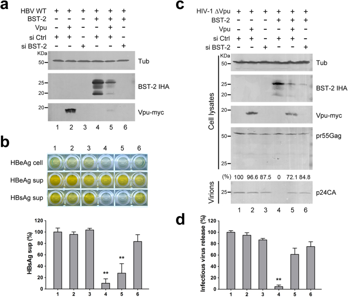
Figure 4. HIV-1 Vpu fails to efficiently inhibit BST-2-induced HBV restriction.
(a) 293T cells were transfected with 1 μg of HBV proviral construct, 50 ng BST-2 IHA, 50 ng Vpu-myc plasmids, and siRNA as indicated. BST-2 IHA, Vpu-myc, and siRNA efficiency were detected by Western blotting. (b) HBV antigen expression and release in a) were examined with ELISA, and HBsAg release percentages are shown. (c) 293T cells were transfected with 1 μg of pNL4-3 ΔVpu proviral construct, 50 ng BST-2 IHA, 50 ng Vpu-myc plasmids, and siRNA as indicated. BST-2 IHA, Vpu-myc, siRNA efficiency, pr55Gag, and concentrated released p24 capsid were detected by Western blotting. (d) The titers of the released infectious HIV-1 viruses in c) were quantified and are shown in percentages. **P < 0.01. These experiments were performed three times, and the most representative Western blot is shown.

