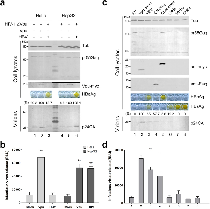
Figure 5. HBV expression enhances HIV-1 ΔVpu release in HepG2 cells.
(a) HeLa and HepG2 cells were co-transfected with 1 μg of pNL4-3 ΔVpu proviral construct, along with 100 ng of empty vector, Vpu-myc vector, or HBV proviral construct. Vpu-myc, cellular pr55Gag, and concentrated released p24 capsid were detected by Western blotting. HBV antigen expression was determined by HBeAg ELISA. (b) The titers of the released infectious HIV-1 viruses in a) were quantified and are shown. (c) HepG2 cells were transfected with 1 μg of pNL4-3 ΔVpu proviral construct, along with 100 ng of empty vector, Vpu-myc vector, or HBV proviral or expression plasmid as indicated. Vpu and HBV core expression was examined by Western blotting with the anti-myc antibody, HBx with the anti-Flag antibody, and the cellular pr55Gag and concentrated release p24 capsid with anti-p24 antibody. HBs and HBe antigen expression was determined by HBsAg and HBeAg ELISA of cultured supernatants. (d) The titers of the released infectious HIV-1 viruses in c) were quantified and are shown. **P < 0.01. These experiments were repeated three times, and the most representative Western blot is shown.

