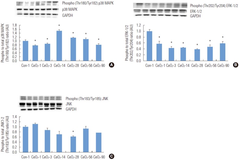
Figure. 5.

Activation of mitogen-activated protein kinase (MAPK) protein signaling was observed with the instillation of cerium oxide (CeO2) nanoparticles. Protein bands of the p38 MAPK, phosphorylated p38 MAPK (A), ERK-1/2-MAPK, phosphorylated ERK-1/2-MAPK (B), and JNK, phosphorylated JNK (C) proteins, along with the corresponding glyceraldehyde 3-phosphate dehydrogenase (GAPDH) levels are represented in the figure bands corresponding to the X-axis labels and are shown in the immunoblotting images. Protein levels were adjusted according to the GAPDH levels and compared with the day 1 control group. One-way analysis of variance was performed for overall comparisons, while the Student-Newman-Keuls post hoc test was used to determine differences between groups. *p<0.05 between the day 1 saline control group.
