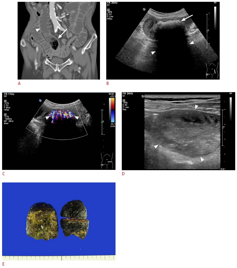
Fig. 1. An 85-year-old man with bezoar-induced small bowel obstruction.

A. Computed tomography shows a mottled gas-patterned intraluminal mass (arrow) suspicious of a bezoar just proximal to the transitional zone of the ileum with the dilated small bowel in front (arrowhead). B. Gray-scale sonogram shows an arc-like surfaced intraluminal mass (arrow) with a strong posterior acoustic shadow (arrowheads). This mass was confirmed as a bezoar after surgery. C. Color Doppler sonogram shows a prominent twinkling artifact (arrowheads) in front of the intraluminal mass. D. Dilated lumen is filled with feces (arrowheads) at the proximal portion of the small-bowel loop. E. The photograph shows the phytobezoar extracted via enterotomy.
