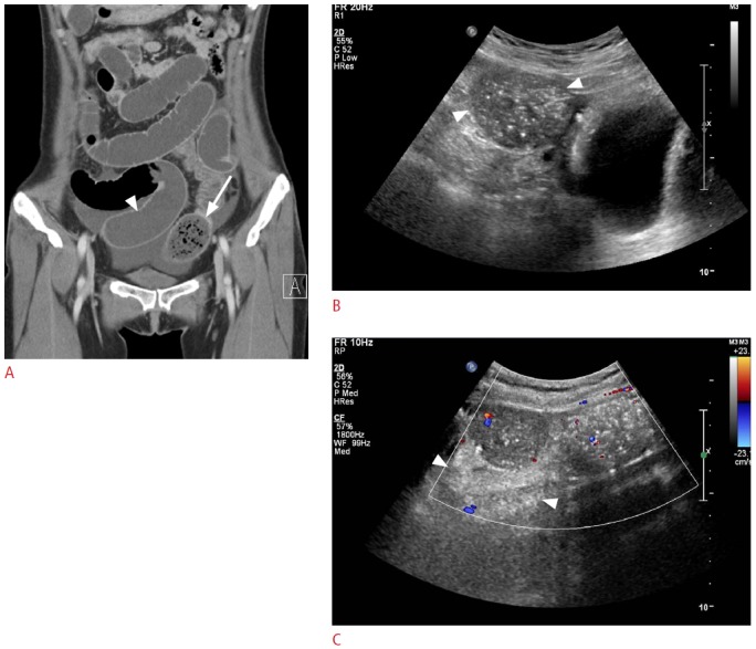
Fig. 2. A 49-year-old woman with small bowel obstruction.

A. Computed tomography (CT) shows a mottled gas-patterned intraluminal mass (arrow) suspicious of a bezoar just proximal to the transitional zone of the ileum with the dilated small bowel in front (arrowhead), which is similar to the findings shown in Fig. 1A. This lesion was clinically diagnosed as feces after spontaneous symptomatic improvement. B. Gray-scale sonogram shows no posterior acoustic shadow or arc-like surfaced mass at the site of lesion corresponding to CT (arrowheads). C. Color Doppler sonogram shows no twinkling around the lesion. Posterior acoustic enhancement (arrowheads) is observed behind the mass-like lesion.
