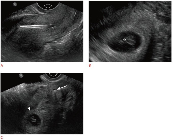
Fig. 6. Displaced intrauterine device (IUD) in a 23-year-old female with positive pregnancy test despite IUD.

A. Sagittal transvaginal sonogram shows malpositioned IUD within the lower uterine segment and cervix. B. An intrauterine pregnancy is seen within the uterine fundus. C. Transverse transvaginal sonogram shows the relationship between the low-lying IUD within the cervix (arrow) and the gestational sac within the uterine fundus (arrowhead). The IUD was removed without incident. The pregnancy resulted in a normal, full-term delivery without adverse complications.
