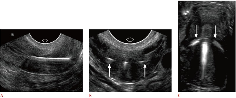
Fig. 9. Embedded intrauterine device (IUD) in a 24-year-old female with menorrhagia and unsuccessful attempts at IUD removal.

A. Sagittal transvaginal sonogram shows inferior displacement of a copper IUD within the cervix. B. Transverse transvaginal sonogram of the cervix shows the arms of the IUD extending into the cervical wall (arrows). C. Three-dimensional coronal sonogram better demonstrates both arms embedded within the myometrium (arrows). The IUD was subsequently removed under anesthesia.
