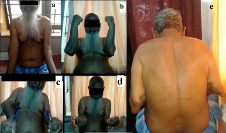
Fig. (2).
Clinical photographs. a) Bilateral elbow joints as viewed anteriorly. b) Bilateral elbow joints with maximal flexion. The prominence of distal humerus and the proximal radio-ulnar complex can be noted on lateral and medial sides respectively. c) Limitation of pronation movement bilaterally. d) Near full range of supination. e) Puckered scars reminiscent of healed sinuses on the posteromedial aspect of elbow.

