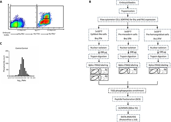
Figure 1. (A) Flow cytometric sorting of ES cells at specific time points of the hematopoietic differentiation for nuclear phosphoproteome analysis.

Differentiating ES cells were enriched based on Bry and Flk1 expression. 2 × 10⁷ cells were used for each experiment. At 2.5 days of differentiation, Bry−Flk1− cells, corresponding to the epiblast-like stage, were collected by flow cytometric sorting (1). At 3.5 days two other populations, Bry+Flk1− and Bry+Flk1+, corresponding to the mesoderm and hemangioblast (BL-CFC) cell populations, respectively, were collected (2). (B) Schematic workflow for the identification of changes in the phosphoproteome of mouse embryonic stem cells committed to the hematopoietic differentiation. Mouse embryonic stem cells sorted for Bry and Flk1 were generated from biological replicates. 100 μg of nuclear lysate were digested with trypsin to produce peptides that were labeled with 4-plex iTRAQTM reagent. Combined samples were then enriched for phosphopeptides via TiO2 column, separated with strong cation exchange chromatography (SCX) and subjected to nano-RPLC-MS/MS on a QTof mass spectrometer. (C) Distribution of the phosphoentity quantification ratios. Data normalization was obtained transforming all control ratios of phosphoentities identified with a sequence confidence above 20% with the logarithmic function at the base 2 (bins ranging from −2 to 2).
