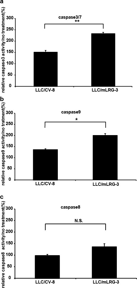
Figure 3. Stimulation with TGFβ1 induced apoptosis more strongly in mLRG-overexpressing LLC cells than in control vector LLC cells.

Caspase-3/7, caspase-8, and caspase-9 activities in LLC/mLRG-3 and LLC/CV-8 cells after treatment with TGFβ1. (a,b,c) Cells were cultivated in 96-well plates and treated with TGFβ1 (1.0 ng/mL) for 24 h. Caspase-3/7 activity was measured with a caspase-3/7 luminescent assay kit (Casapase-GloTM). Similarly, caspase-8 and caspase-9 activities were measured using a caspase-8 and caspase-9 luminescent assay kit (Casapase-GloTM). Each relative value (TGFβ1 treatment/no treatment) is the average ± standard deviation. * P < 0.05, **P < 0.01.
