
Figure 1. Kinetics of vascular channel formation in HUVEC, MDA-MB-231, and MCF-7 cells.
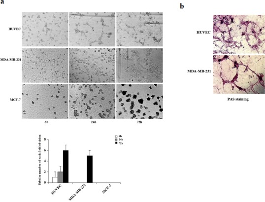
(a) HUVEC, MDA-MB-231, and MCF-7 cells were plated on matrigel, and images were acquired using phase-contrast microscopy at the times indicated. HUVEC cells started to form vascular channels 6 h after plating whereas MDA-MB-231 cells formed vascular channels at 24 h. Well-defined patterned networks were observed by 72 h. MCF-7 cells failed to form patterned networks. (b) Periodic Acid-Schiff (PAS) staining of HUVEC and MDA-MB-231 cells plated on matrigel for 72 h to identify secreted extracellular matrix. Pink staining identifies glycogen and related mucopolysaccharides secreted by cells to form the extracellular matrix-rich vascular channels.