
Figure 3. Effects of claudin blocking antibodies on cell proliferation, morphology, and tubule formation.
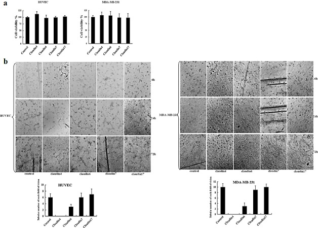
(a) MTT assay was used to assess the effects of different blocking antibodies on the proliferation of HUVEC and MDA-MB-231 cells. HUVEC and MDA-MB-231 cells were plated in 96-well plates. Medium containing claudin-2, -3, -4, -6, -7, or -17 blocking antibodies (1 μg/mL) were added to the 96-well plates. An equivalent volume of mouse IgG1 control antibody was used as a control. Data represent the mean + SD (n=3). *: p < 0.05 compared with controls. (b) HUVEC and MDA-MB-231 cells were also cultured in matrigel. Images were acquired by phase-contrast microscopy at the times indicated.