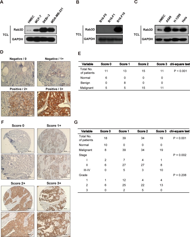
Figure 1. Increased Rab3D in malignant tumor cells and in clinical cancer specimens.
(A). Western Blot analysis of intracellular Rab3D expression in HMEC, non-invasive breast cancer MCF-7, and invasive SKBr-3, MDA-MB-231 cell lines. (B). Western Blot analysis of intracellular Rab3D expression in melanoma F0, F1, F10 cell lines. (C) Western Blot analysis of intracellular Rab3D expression in lung cancer A549, H-1299 cell lines and ovary cancer Hela cell line. (D). Representative Rab3D staining in normal tissues, benign tumor and malignant breast cancer samples that illustrate immunohistochemical scores of 0, 1, 2 and 3. Scale bar, 100 μm. (E). Association of Rab3D immunohistochemical scores with tumor malignancy. The x2 test was used to test difference between categorical variables. (F). Representative Rab3D staining in different stages of malignant breast cancer samples that illustrate immunohistochemical scores of 0, 1, 2 and 3. Scale bar, 100 μm. (G). Associations of Rab3D immunohistochemical scores with tumor stage (I, II, III-IV) and tumor grade (1, 2, and 3). The x2 test was used to test difference between categorical variables.

