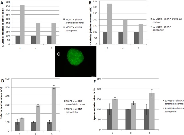
Figure 3. Silencing of spinophilin increases anchorage-independent growth and tumor sphere formation.

Graphs represent the results from three independent biological replicates of the breast cancer cell lines MCF-7 (A) and SUM159 (B) stably transfected with shRNA against spinophilin compared to control cells. In each biological replicate, a significantly increased number of colonies were observed in the spinophilin-silenced cells. (C) A representative example of a tumor sphere (mammosphere) under ultra-low attachment conditions. The transfected cells are labeled with green-fluorescent protein. Significant increase in the number of mammospheres after spinophilin-silencing in MCF-7 (D) and SUM159 (E) cells.
