
Figure 1. Characterization and differentiative properties of CSC from breast and renal carcinomas.
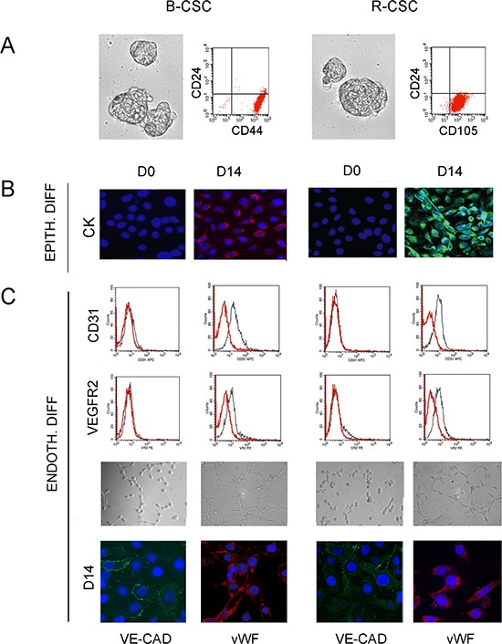
Panel A and B. B-CSC and R-CSC grew in spheres and were characterized as CD24−/CD44+ or CD24−/CD105+ cells, respectively (A). B-CSC and R-CSC lacked cytokeratin (CK) that was acquired when cultured in epithelial differentiating conditions (EPITH. DIFF.) for 14 days (D14), as compared with basal condition (D0) (B). Panel C. B-CSC and R-CSC cultured for 14 days (D14) in endothelial differentiating conditions under hypoxia (ENDOTH. DIFF.) acquired the endothelial-specific markers CD31, VEGFR2, VE-cadherin (VE-CAD) and vWF and the ability to organize into capillary-like structures. Original magnification: immunofluorescence staining: x400; tubulogenesis: x200. Nuclei were counterstained with Hoechst dye.