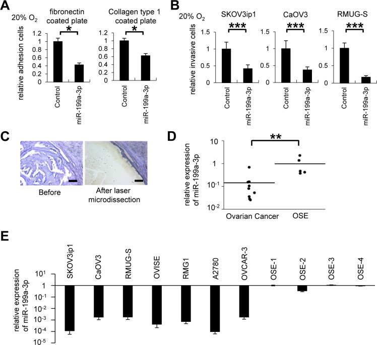
Figure 2. MiR-199a-3p displays tumor suppressor functions, and its expression is downregulated in ovarian cancer specimens and cell lines.
(A) In vitro adhesion assay. SKOV3ip1 cells (5 × 10⁴ cells) were plated onto fibronectin- or collagen type 1–coated 96-well plates under 20% O2 for 75 minutes. Data represent mean ± SEM, n = 5. (B) Matrigel invasion assay. Ovarian cancer cells (5 × 10⁴ cells) were plated in a Matrigel-coated upper chamber in serum-free medium and allowed to invade the lower chamber, which contained in 10% fetal bovine serum + Dulbecco's modified Eagle's medium for 24 hours under 20% O2 condition. Data represent mean ± SEM, n=7. (C) Representative images of selective collection from formalin fixed paraffin-embedded tissues of ovarian cancer tumor region (high-grade serous adenocarcinoma) using laser capture microdissection. Bar represents 200 μm. (D) miRNA RT-qPCR. Ovarian cancer tumor tissue (high-grade serous adenocarcinoma) or normal ovarian surface epithelium was selectively collected. The expression of miR-199a-3p was significantly downregulated in ovarian cancer clinical samples. (E) miRNA RT-qPCR. The expression of miR-199a-3p in 7 ovarian cancer cell lines (SKOV3ip1, CaOV3, RMUG-S, OVISE, RMG1, A2780, and OVCAR-3) was significantly lower than that of OSE (ovarian surface epithelium); *P < 0.05; **P < 0.01; ***P < 0.001.

