
Figure 3. Uptake of OP9 exosome content by ALL cells.
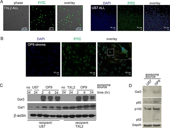
A, B. Confocal microscopy of leukemia or stromal cells incubated with labeled heterologous exosomes. A: Human TXL2 ALL or US7 cells with OP9 exosomes. B: OP9 stromal cells with US7 exosomes. Bar, 20 μm (panel A-TXL2) or 16 μm (panel A-US7 and panel B). C. Western blots of lysates from US7 or TXL2 ALL cells cultured without OP9 stroma for 24 hours, then exposed to 50 μg/ml stromal or “self” ALL exosomes for 2–24 hours in serum free medium. 20 μg protein/lane. One of two independent experiments with similar results. D. Western blot of lysates (1.5 × 10⁵ cell equivalent/lane) from US7 cells cultured without OP9 or serum, then stimulated for 24 hours with 0.5 μg/μl US7 or OP9 exosomes. Antibodies used as indicated to the left.