
Figure 6. Chemotherapeutic drug treatment induces endogenous Galectin-3 in pre-B ALL cells when in contact with stromal cells.
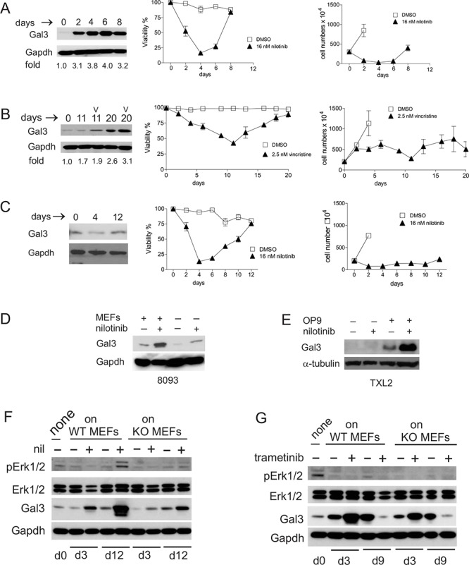
A. Left panel, Western blot analysis for Galectin-3 expression; middle panel, viability and right panel, cell counts of murine Bcr/Abl-positive 8093 pro/pre-B ALL cells treated with nilotinib. Western blot: fold increase is with respect to Galectin-3 levels on d0. Cell counts: cell numbers are not indicated for all days in the control samples since removal of cells was necessitated by overcrowding due to exponential cell growth. B. Human US7 ALL cells treated with 2.5 nM vincristine (v) in the presence of irradiated OP9 cells. Panels as in A. C. Murine 8093 ALL cells treated with 16 nM nilotinib but without direct contact with MEFs. Panels as in A. D. 8093 cells treated with or without 16 nM nilotinib for 48 hours in the presence or absence of MEFs. E. Human Ph-positive TXL2 cells treated with or without 1 μM nilotinib for 72 hours in the presence or absence of OP9 cells. F, G. 8093 pro/pre-B ALL cells cultured without MEFs for 24 hours (input d0 samples designated ‘none’), then plated on gal3+/+ [WT] or −/− [KO] MEFs as indicated above the panels, were harvested on different days of treatment with 16 nM nilotinib (F) or 10 nM trametinib (G) Antibodies used for Western blots are shown to the left in each panel. Gapdh, loading control. F, representative image, one of two independent experiments. In all experiments, fresh drug was added with each fresh medium change.